
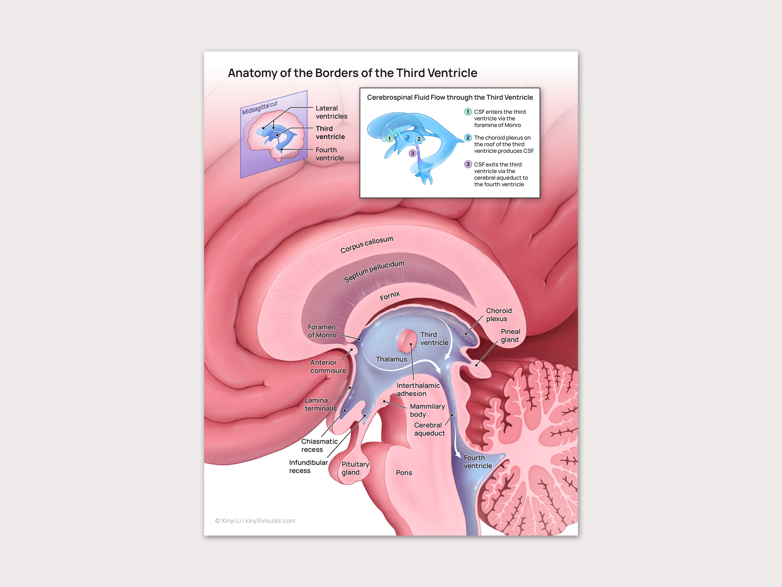
Anatomy of the Third Ventricle
Neuroanatomy • 2022
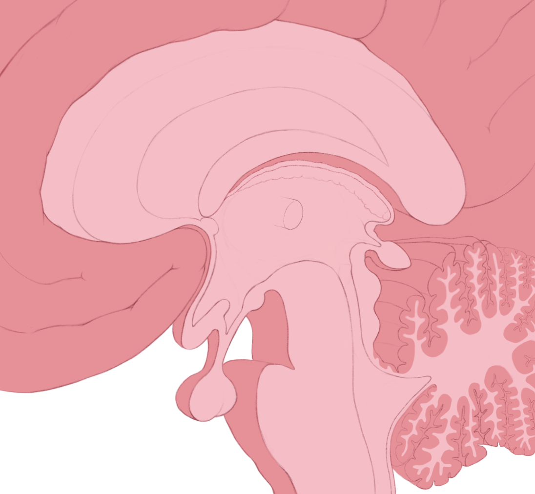
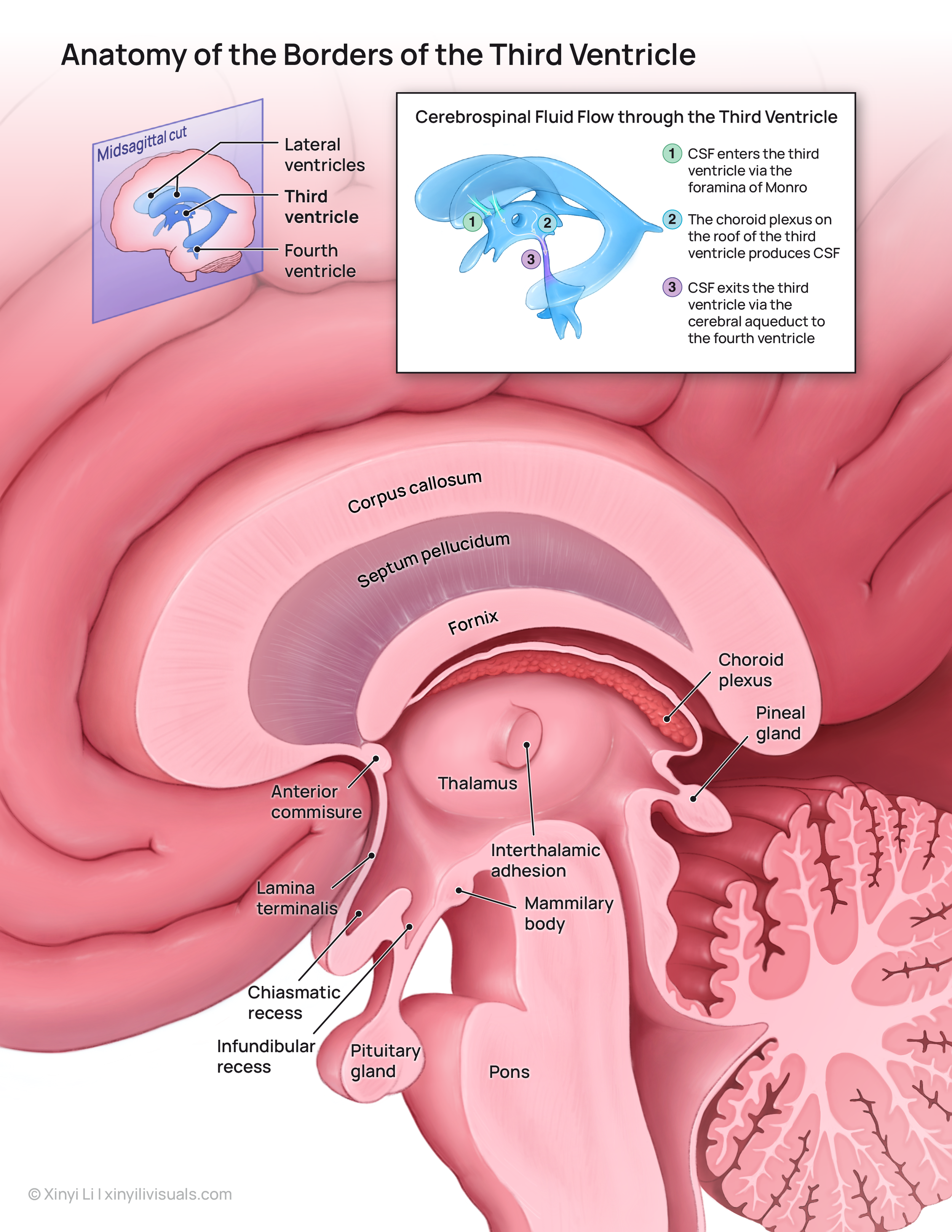
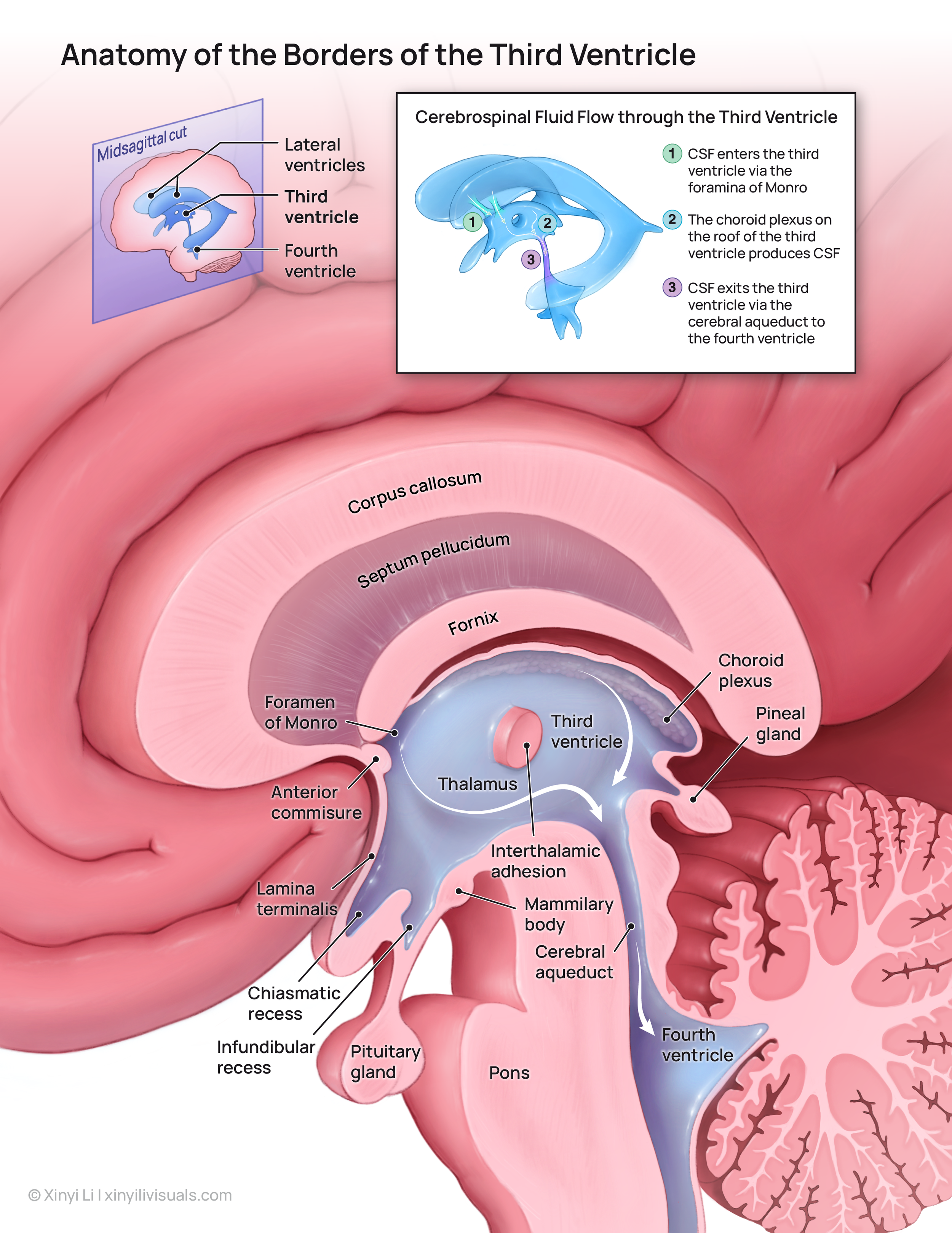
A didactic illustration and interactive slider aimed to educate neuroanatomy students on the anatomy and function of the third ventricle. In particular, the structures of the deep brain that form its borders and the direction of CSF flow through the third ventricle.
Client
Dr. Shelley Wall
Software
Adobe Illustrator
Procreate
Github
Format
Didactic illustration
Interactive slider
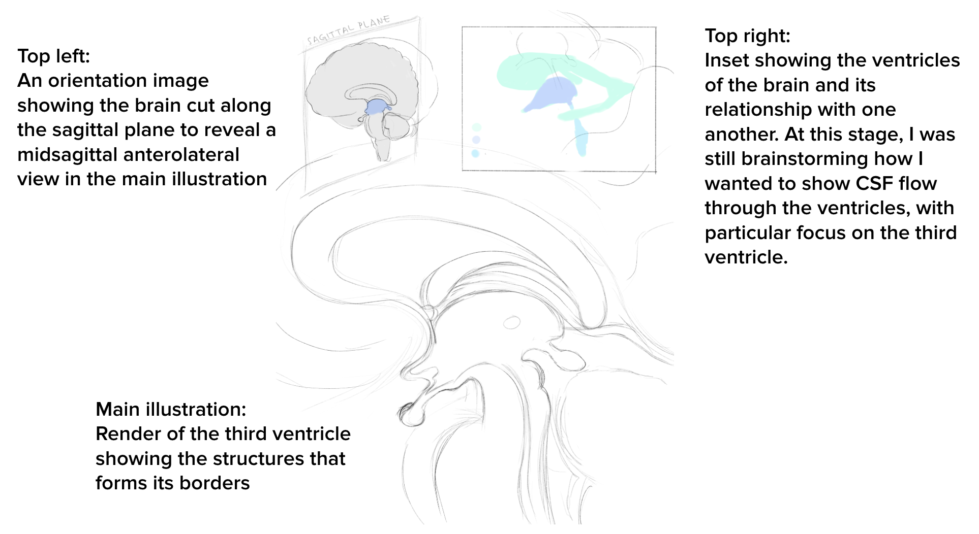
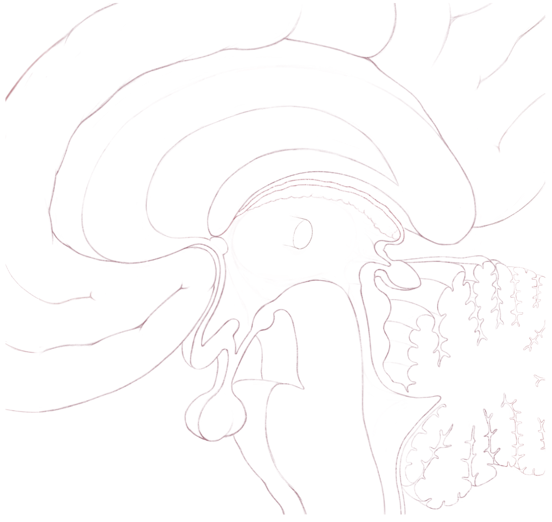
Ideation & Rough Sketch
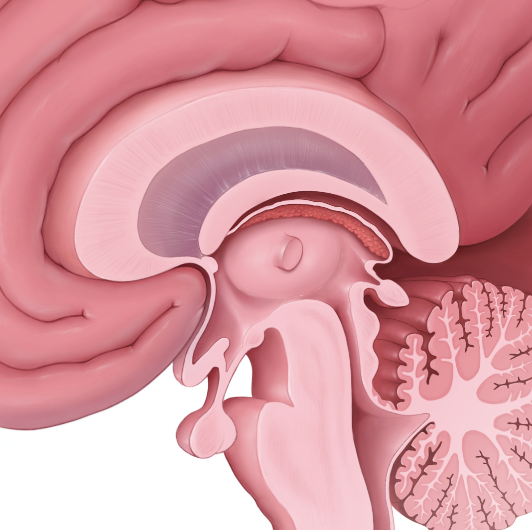
The original idea for this assignment began with a conversation I had with a classmate, who shared the sentiment that it was difficult to conceptualize at first that the ventricles of the brain were empty spaces since they are so often depicted as an actual structure inside the brain.
I decided to focus on depicting the structure of the third ventricle, as it is surrounded by many structures of the deep brain which form its walls. After consulting with Dr. Wall, I was inspired to incorporate CSF flow to and from the third ventricle into the illustration as well, since it would give more insight into the function of the third ventricle itself, and also give structural context on how it connects to the lateral and fourth ventricles.
Research
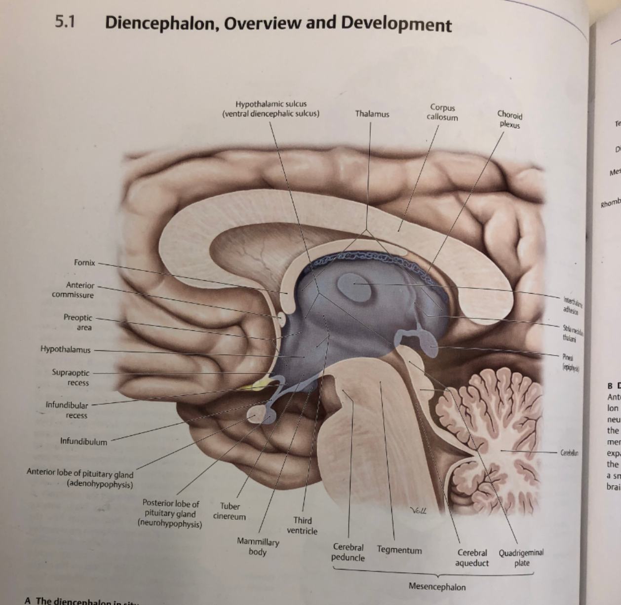
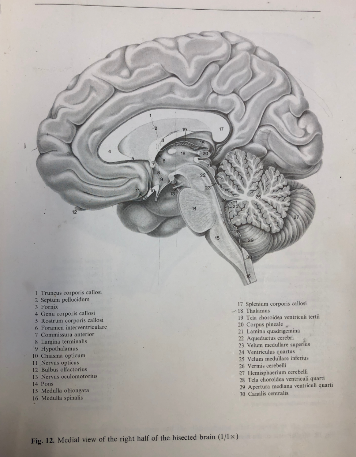
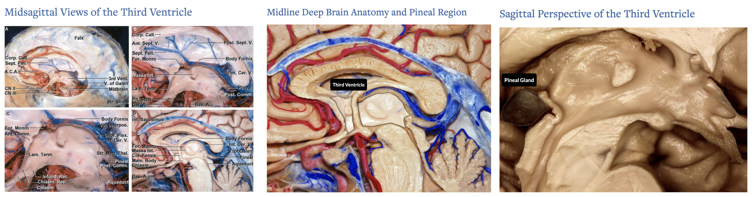
I referenced textbook illustrations as well as articles from the Neurosurgical Atlas to understand the anatomical relationship of the structures within the deep brain.
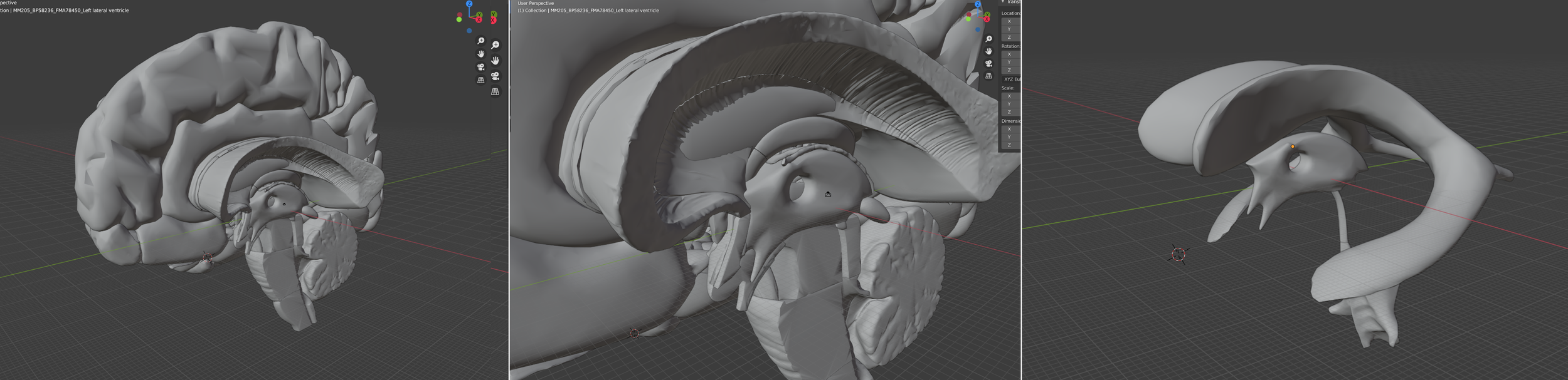
I created a maquette based on data from https://lifesciencedb.jp/bp3d/?lng=en , then oriented the maquette in the angle that I wanted to illustrate from which I would be able to trace to get a general sense of the anatomical shape & structures.
Production
REFERENCES
Agur, A., Dalley, A., & Boileau, G. (2009). Grant's Atlas of Anatomy. Lippincott Williams & Wilkins.
Schünke Michael, Schulte, E., & Schumacher, U. (2007). Head and neuroanatomy: Thieme Atlas of anatomy. Thieme.
https://www.neurosurgicalatlas.com/neuroanatomy/midsagittal-views-of-the-third-ventricle
https://www.neurosurgicalatlas.com/neuroanatomy/midline-deep-brain-anatomy-and-pineal-region
https://www.neurosurgicalatlas.com/neuroanatomy/sagittal-perspective-of-the-third-ventricle
https://www.neurosurgicalatlas.com/volumes/brain-tumors/intraventricular-tumors/anatomy-of-the-ventricular-system
https://www.neurosurgicalatlas.com/neuroanatomy/floor-and-roof-of-the-third-ventricle-2
https://sketchfab.com/3d-models/the-third-ventricle-2c540ae3a2704ebdb603dcdab8793744
https://lifesciencedb.jp/bp3d/?lng=en