Neuroanatomy Portrait
Neuroanatomy • 2022
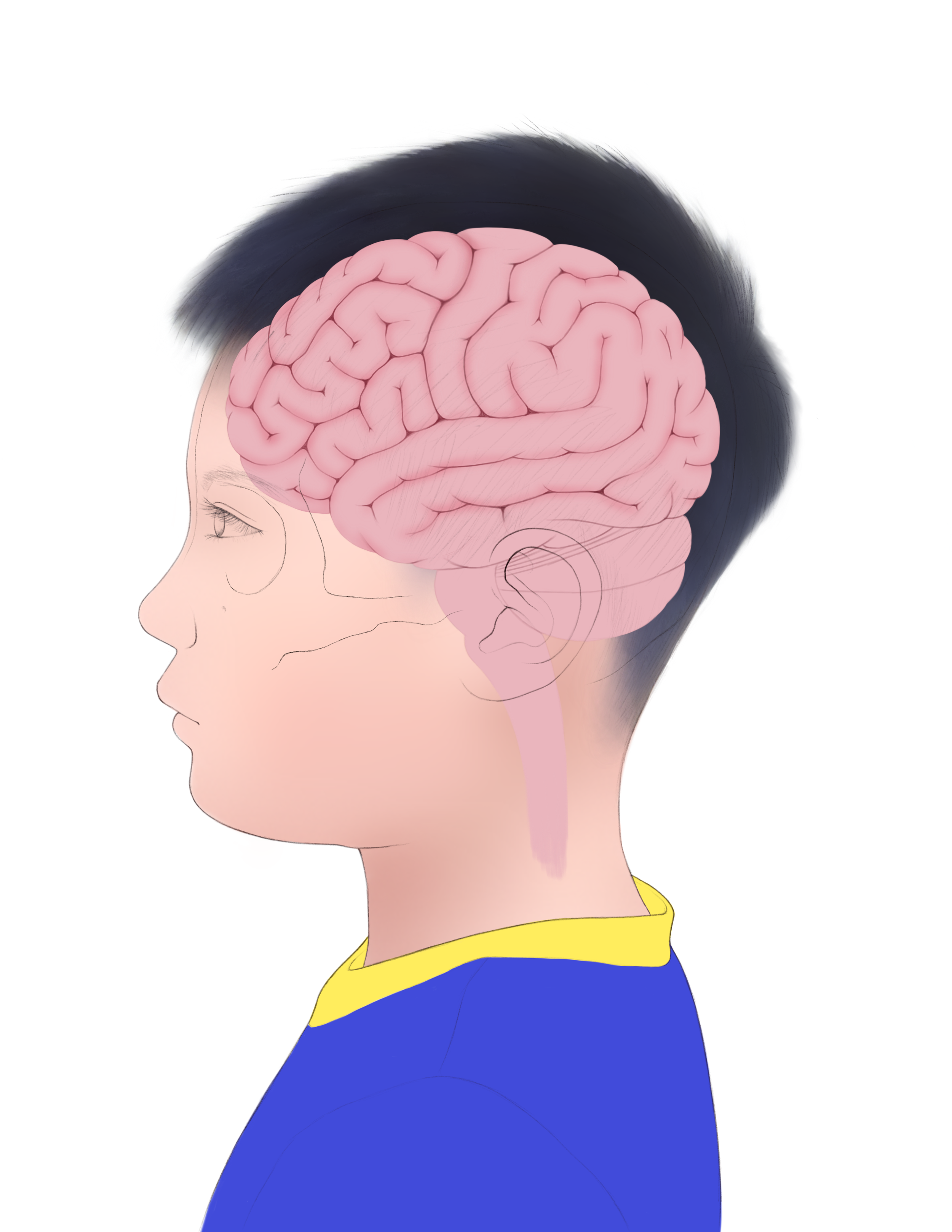
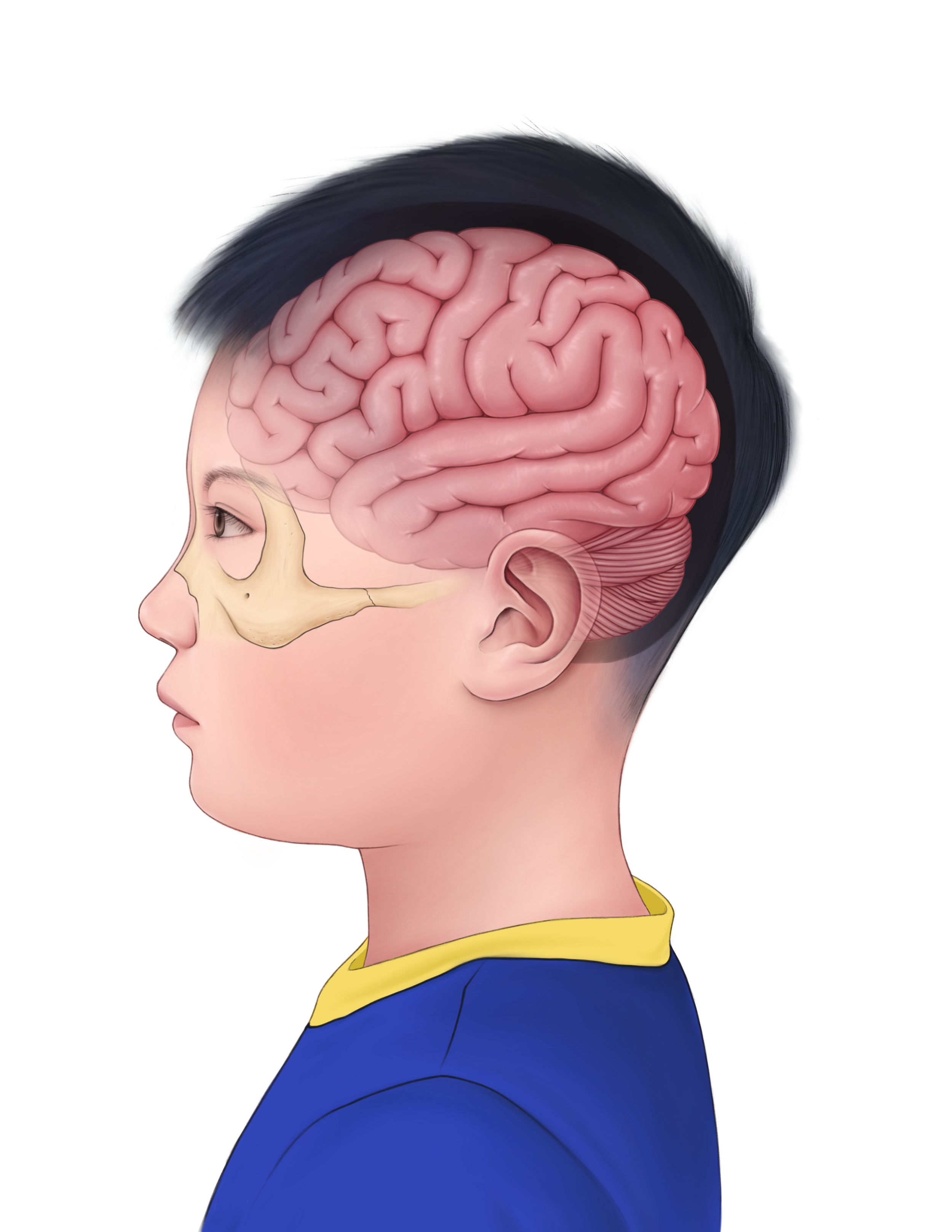
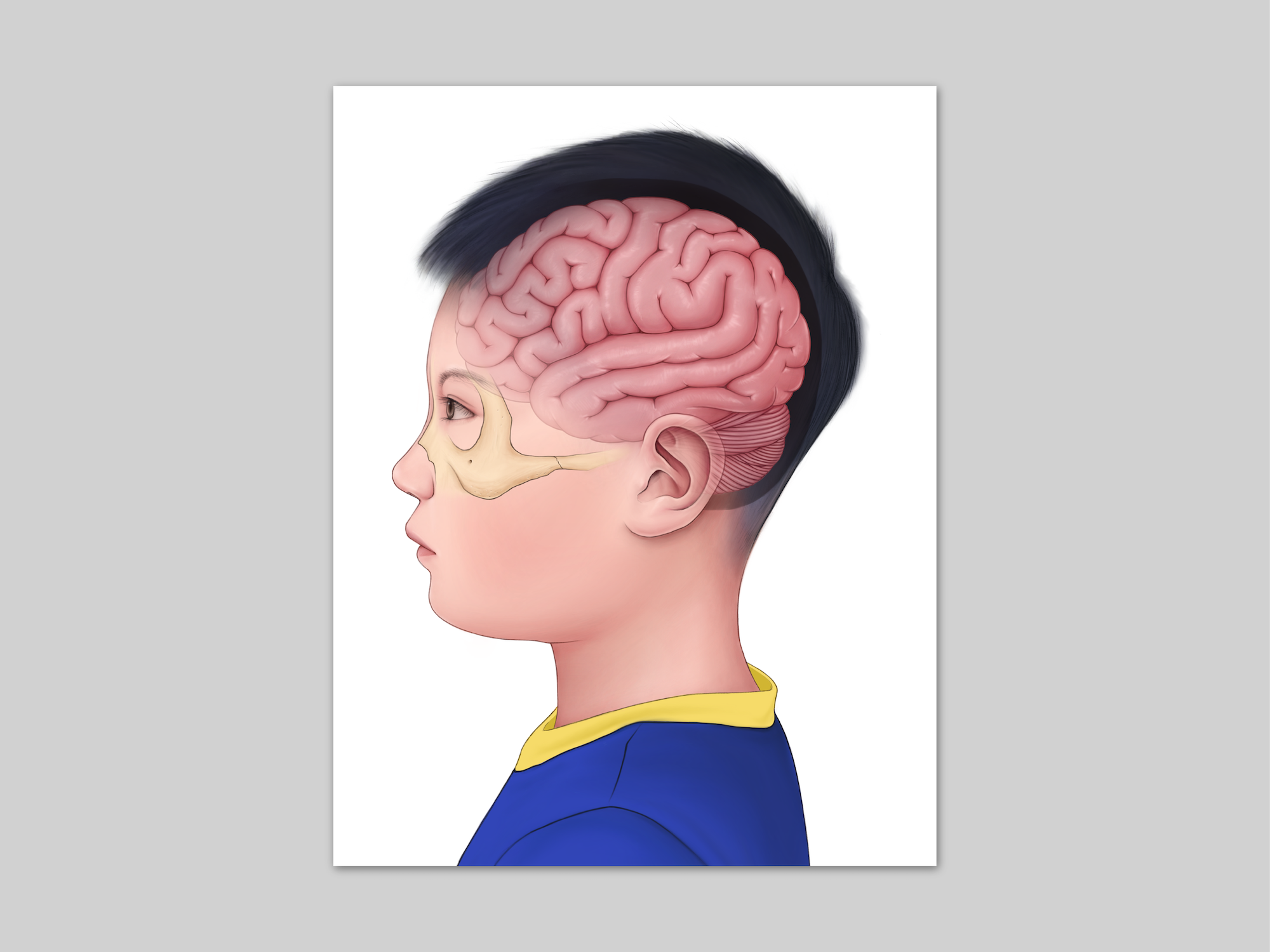
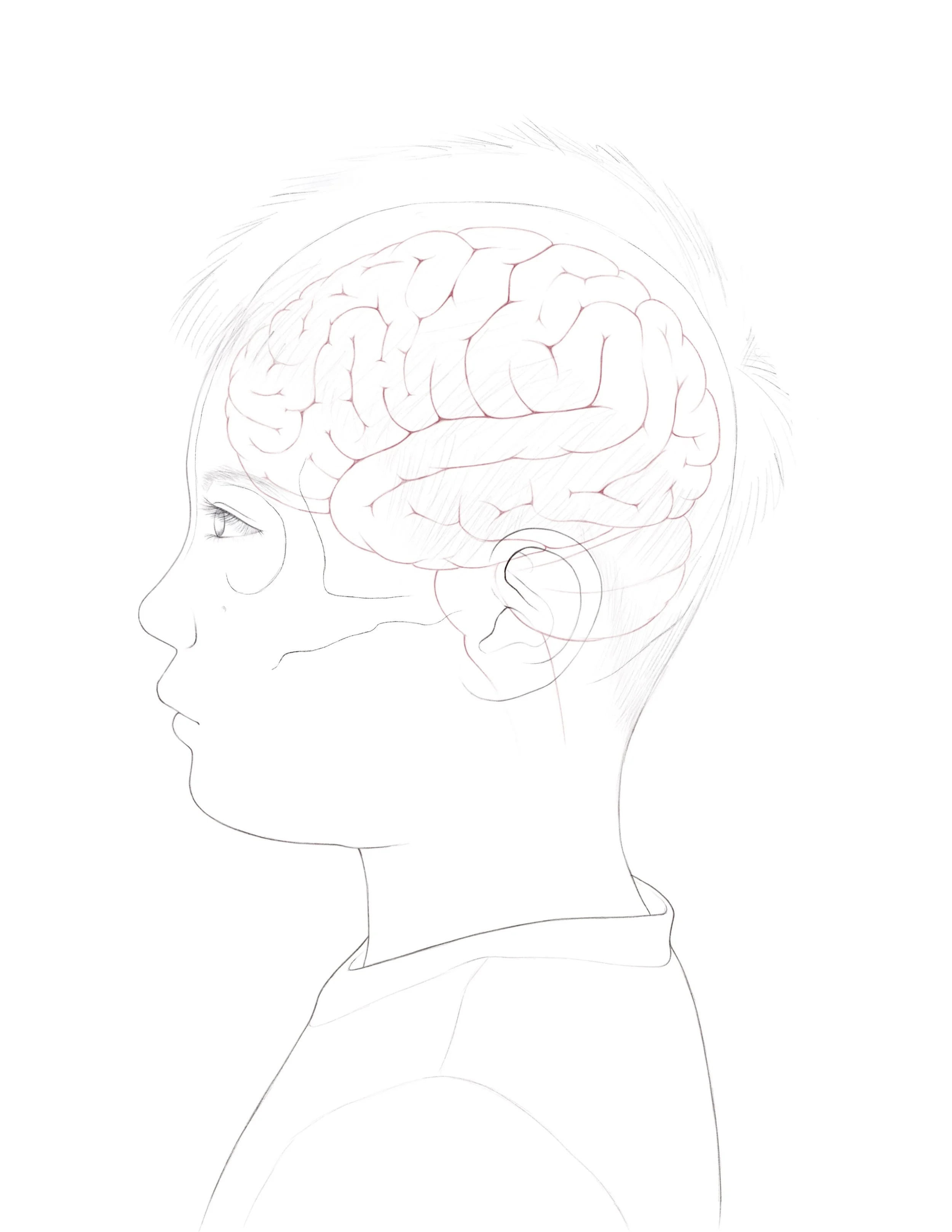
An editorial illustration created to accurately visualize the gross anatomy of the brain in relation to the cranial cavity.
Client
Dr. Shelley Wall
Software
Adobe Photoshop
Adobe Illustrator
Procreate
Format
Illustration
Poster
Ideation
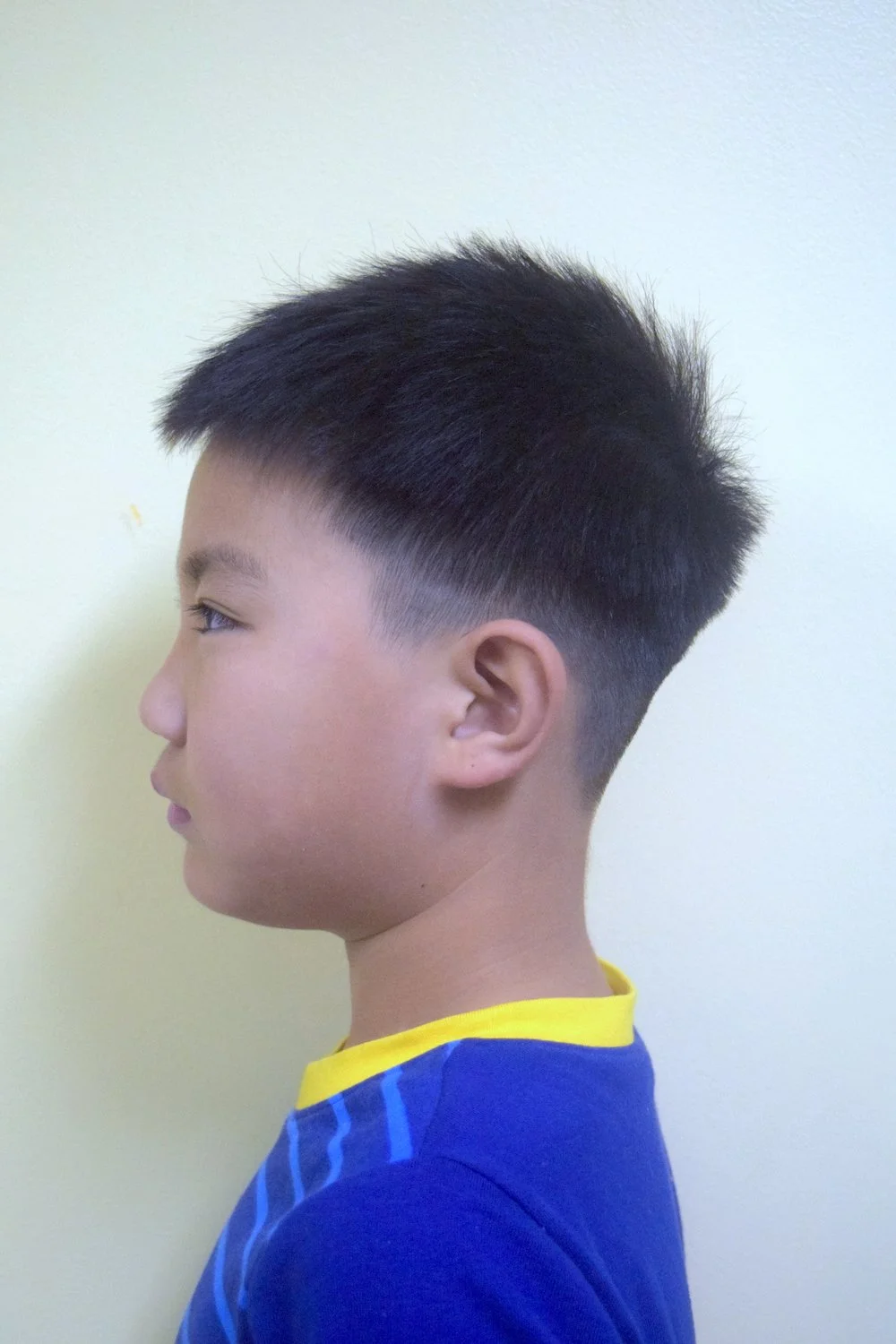
The process for this illustration began with taking a reference photo to orient the skull and brain. I chose to illustrate my brother and took photos of him from a lateral view.
As I chose to illustrate a portrait of my brother, I wanted to portray the childhood brain in a gentle, illustrative style that would be appropriate for patient education. I was inspired by an illustration from Mayo Clinic, which utilizes a soft pencil-like rendering style and places visual emphasis on the brain by employing more saturated colours on the brain compared to the rest of the illustration.
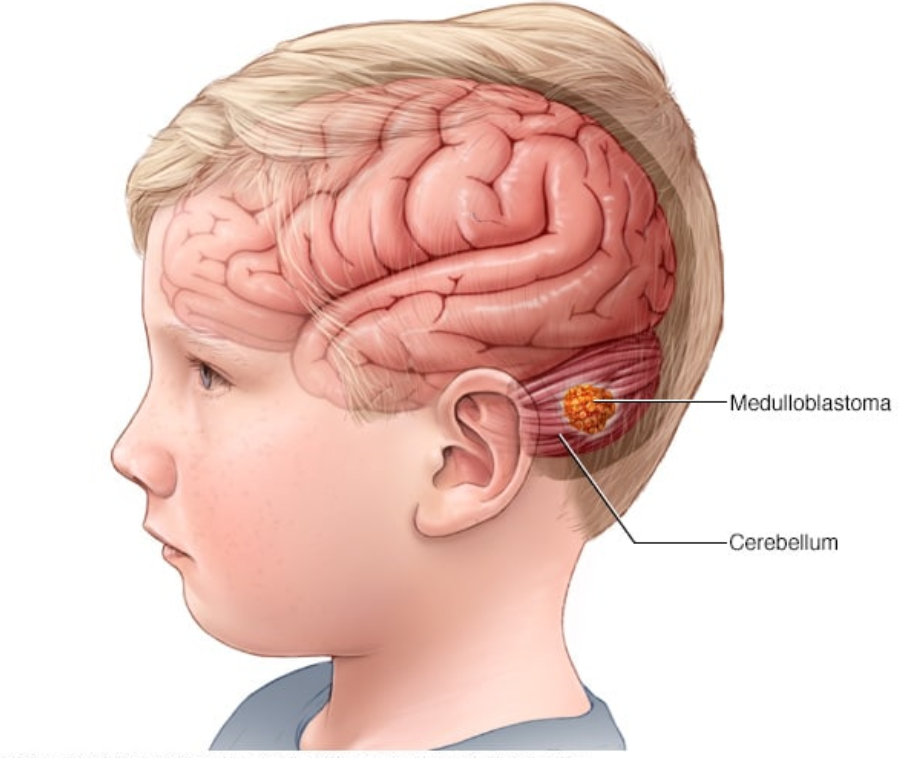
Mayo Clinic Staff. (2021, August 6). Brain Tumor. Mayo Clinic. Retrieved October 6, 2022, from https://www.mayoclinic.org/diseases-conditions/medulloblastoma/cdc-20363524
Research & Reference Images
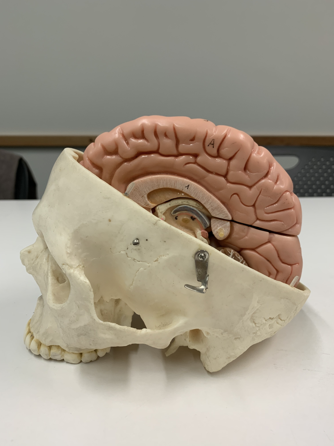
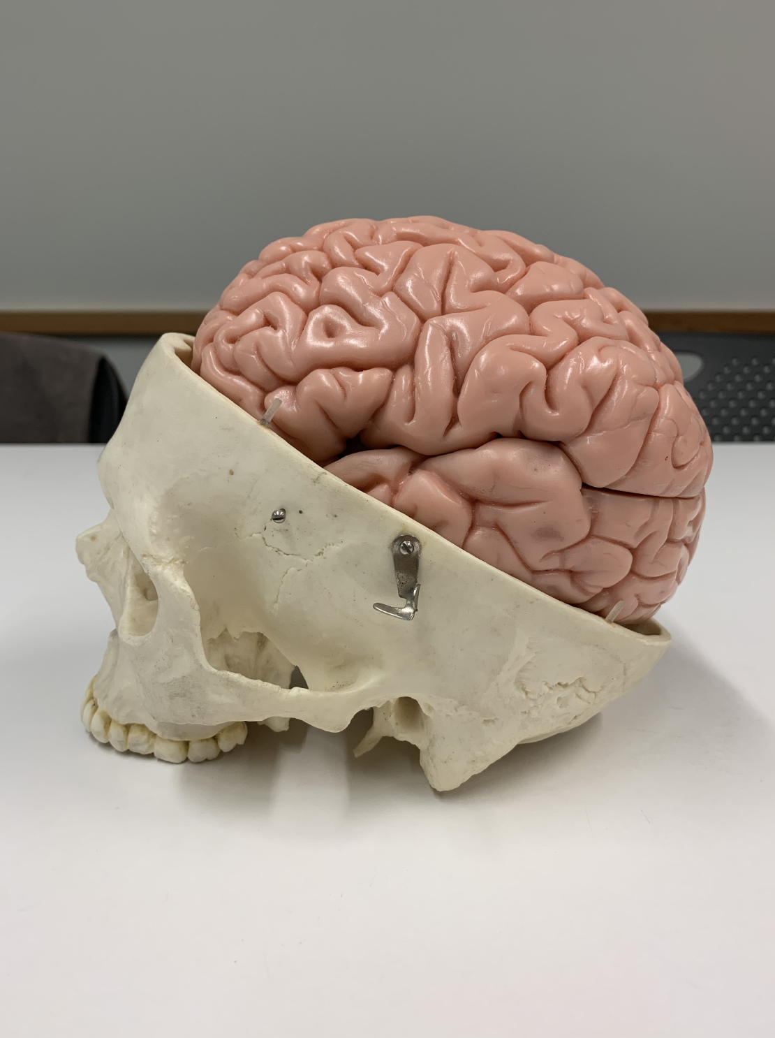
I took reference images from models, which were helpful for orienting the brain inside the skull.
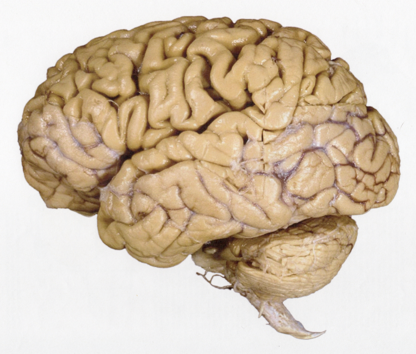
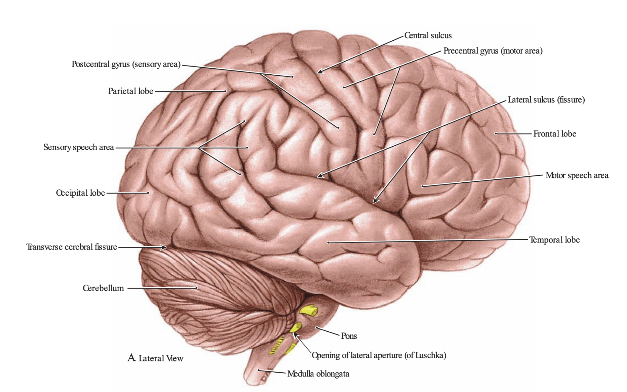
Agur, A. M. R., & Dalley, A. F., II. (2017). Neuroanatomy: Overview and Ventricular System. In Grant's atlas of anatomy. (14th ed., pp. 698-701). Philadelphia, Wolters Kluwer.
Grant’s atlas was my primary reference for the brain’s surface anatomy. I also referenced plastinated images provided by Dr. Wall, taken from Gray’s Anatomy.
Comprehensive Sketch to Final Render